1999-highton-43

Highton J, Guévremont D, Thomson J, Carlisle B, Tucker I. A trial of clodronate-liposomes as anti-macrophage treatment in a sheep model of arthritis.Clin. Exp. Rheumatol. 1999 Jan;17(1):43–8.

Abstract

Objective
Our previous research has concerned the role of macrophages in joint inflammation in rheumatoid arthritis. We have therefore been interested in liposomes containing clodronate as an anti-macrophage treatment for arthritis. We have used the antigen-induced arthritis model in sheep to evaluate the effect of clodronate liposomes.

Methods
Arthritis was induced in the right hock joint (day 0). We were able to demonstrate uptake of liposomes into macrophages within the inflamed joint lining. On day 7, sheep were given a single intra-articular injection of clodronate liposomes (group 1, n = 10) or saline liposomes (group 2, n = 10). A further 6 sheep (group 3) had no arthritis and no treatment.

Results
No difference in joint diameter was observed between the sheep in group 1 (clodronate) and group 2 (saline treated). Both groups had joint swelling which persisted until the end of the trial (day 20). Histologic scoring was also similar in group 1 and group 2 animals, and both were worse than group 3.

[custom_table]
Clodronate Concentration Total Lipid Concentration Lipid Composition Lipid Mole % Liposome Type Control Liposomes Control Free Clodronate
not specified (see pt. 1) 42 mg/ml EPC/BPS/chol 48/48/4 MLV saline ND
[/custom_table]  [custom_table]
Animal Description Clodronate Dose Dosing Method/Site Target Phagocytes Systemic Dosing? Systemic Results
Sheep 0.5 ml intra-articular/knee synovial no N/A
[/custom_table]

Notes

  1. Liposome prep method cited – none.
  2. We calculated the lipid concentration assuming that no lipid was lost during the preparation of the liposomes; this is a tenuous assumption.
  3. The author’s stated that the clodronate liposomes were unilamellar, but no size information was provided. While probe sonication can provide small unilamellar liposomes in the range of 50 nm or smaller with this lipid composition, the key is to verify that the liposome suspension converts from an opaque, milky suspension to a translucent suspension which indicates that no large multilamellar liposomes remain in the suspension. The sonication time (5 minutes in this case) required to achieve small unilamellar vesicles (SUV) will vary with volume, vessel geometry, sonicator output, etc., therefore cannot be used to verify that small unilamellar vesicles (SUV) were achieved. The authors did state that the unencapsulated clodronate was removed by centrifugation with no further details. Small unilamellar liposomes are very difficult to pellet by centrifugation but it can be done using an ultracentrifuge and extended centrifugation times of several hours at 100,000xg. If the liposomes are truly SUV, low speed centrifugation (10,000xg range) that is used for multilamellar clodronate liposomes will provide a barely dectable, if present at all, pellet.
  4. Assuming that small unilamellar liposomes were prepared by the authors as they implied, the problem with SUV is that they encapsulate a very small volume of aqueous phase due to their small internal volume. The aqueous volume of SUV is around 0.5 µl/µmole of lipid or less. The authors reported using 217 µmoles of lipid (by our calculations) which would mean that 108.5 µl of clodronate solution was encapsulated. Again, assuming that the clodronate solution used to rehydrate the dry lipid film was not diluted during the preparation and washing of the liposomes (this is rarely true, and is almost certainly not true in this case since the clodronate solution is hyperosmotic to the saline used for washing the liposomes), then a maximum of 21.7 mg of clodronate would have been encapsulated in the 2 ml clodronate SUV suspension. The authors stated that 200 mg/ml clodronate in liposomes was dosed at 0.5 ml, or 100 mg clodronate, per knee joint; by our calculations, a maximum of 5-5.5 mg of clodronate could have been dosed, if the authors did indeed prepare clodronate SUV-type liposomes. It is very important to characterize the liposome preparations used for treatments at least by assaying the final concentrations of lipid and clodronate in the preparation, yet this is rarely reported in the clodronate liposome literature. Therefore, as in this case, we have no idea of how much clodronate was inside the liposomes that the animals received. We usually make assumptions based on “typical” clodronate liposome preparations, but the liposomes described in this paper are not typical clodronate liposomes.
  5. Antigen-induced arthritis (AIA) was primed by 3 immunization injections of 10 mg ovalbumin (OVA) emulsified in Freund’s Complete Adjuvant (FCA) at 2 week intervals.
  6. Delayed-type hypersensitivity (DTH) for OVA was assessed 24-48 hr post-antigent injection to ensure a response to OVA.
  7. AIA was induced by intra-articular injections of 5 mg OVA in 0.5 ml into the right hocks of sheep while the left (control) hock received no injection.
  8. 7 days later, intra-articular injections of 0.5 ml clodronate liposomes, saline liposomes or steroid(s) were made into the arthritic joints.
  9. BODIPY-PC-containing liposomes were injected into arthritic hocks to assess the uptake of liposomes into the synovial lining of the joints; no details of liposome prep or dose volume was provided. We assume that the authors prepared them by the same method as used for preparing the control liposomes, but this was not stated in the paper.

Results

  1. The authors chose the sheep AIA model because the size of the sheep hocks would allow for more accurate intra-articular injections and joint measurements.
  2. The goal of these experiments was to evaluate the efficacy of liposomal clodronate in the treatment of antigen-induced arthritis (AIA) in sheep, yet the authors did not state the dose of liposomal clodronate that was tested in this model. A 0.5 ml volume of an unknown concentration of liposomal clodronate was dosed intra-articularly to sheep one time at 7 d post-induction of AIA.
  3. No response was seen with either clodronate or control liposomes with respect to joint swelling.
  4. The histological analysis seemed to suggest that both clodronate and control liposomes worsened the damage to the joints when compared to untreated arthritic joints.
  5. When a volume of clodronate liposomes was incubated in heparinized sheep blood ex vivo, the authors reported that liposomes were taken up by monocytes with maximal uptake after 4 h (data not shown).
  6. When a volume of clodronate liposomes was added to cultured macrophages, the authors reported that macrophages died (data not shown).
  7. When a volume of clodronate liposomes or control liposomes was added to a monocyte suspension, a respiratory burst was observed and the authors concluded that liposomes caused a pro-inflammatory response.
  8. When a volume of clodronate liposomes was added to a PMN suspension, similar results were observed leaving us to wonder if the PMN actually endocytosed the clodronate liposomes. Studies in which PMN are incubated with clodronate liposomes usually do not result in the liposomes being taken up by PMN, although there are several reports in the literature of liposome uptake by PMN. However the liposomes prepared in the Highton, et. al. lab may be much smaller than multilamellar clodronate liposomes and contain phosphatidylserine (used only in a few other clodronate liposome studies). Details of the nature of the effect of the clodronate liposomes on PMN which resulted in a respiratory burst would have been a unique contribution to the literature, but…data not shown.
  9. When a volume of fluorescent liposomes was injected intra-articularly into arthritic sheep hocks, the authors reported that confocal microscopic analysis of the synovial lining at 2, 6 and 24 h indicated that labeled synovial macrophages do take up fluorescent liposomes, however our copy of the paper does not include a colored image for confirmation. The authors further stated that uptake was maximal at 6 h and that “Liposomes had dispersed by 24 hours.”
  10. Given that this was an atypical clodronate liposome formulation which was not characterized post-preparation, we find little useful data in this report which is unfortunate considering the cost limitations of performing experiments in large animal models.

 

[divider_top]

2001-čeponis-1908

Čeponis A, Waris E, Mönkkönen J, Laasonen L, Hyttinen M, Solovieva SA, Hanemaaijer R, Bitsch A, Konttinen YT. Effects of low-dose, noncytotoxic, intraarticular liposomal clodronate on development of erosions and proteoglycan loss in established antigen-induced arthritis in rabbits. Arthritis Rheum. 2001 Aug;44(8):1908–16.

Abstract

OBJECTIVE
To assess the clinical and histologic effects of an intraarticular application of low-dose (non-cytotoxic) liposomal clodronate in established antigen-induced monarthritis (AIA) in rabbits.

METHODS
AIA was monitored by assessments of joint swelling, C-reactive protein levels, and radiographic changes in 17 NZW rabbits for 8 weeks during the course of weekly intraarticular injections of liposomal clodronate (0.145 mg/injection, low dose) or “empty” liposomes. The contralateral knee was injected with liposome buffer alone as the control. End-point analyses included macroscopic joint examination, immuno- and TUNEL staining, Safranin O staining/microspectrophotometry, and tumor necrosis factor alpha (TNFα) convertase enzyme (TACE) inhibition assay.

RESULTS
Liposomal clodronate-treated rabbits showed a reduction and delay in joint swelling during the first 3 injections. Expression of matrix-bound (solubilized) TNFα, lining cell hyperplasia, and levels of RAM-11+ macrophages were low in the synovium of the liposomal clodronate treatment group, but the proportion of apoptotic lining cells was not affected. The radiologic score was low at the end of weeks 2 and 4, but at 8 weeks, no difference, compared with controls, was found in pannus formation or in the extent of joint erosion; also, joint swelling was higher than before initiation of treatment. Injections of liposomal clodronate prevented cartilage proteoglycan loss, which was significant in the superficial zone only. TACE activity was not inhibited by clodronate.

CONCLUSION
Liposomal clodronate had temporary antiinflammatory and antierosive effects on established AIA in rabbits. Over the long-term, the loss of cartilage proteoglycans was halted. This observed treatment effect may be related to the inhibition of TNFα production and processing in the synovium.

[custom_table]
Clodronate Concentration Total Lipid Concentration Lipid Composition Lipid Mole % Liposome Type Control Liposomes Control Free Clodronate
0.29 mg/ml Not spec. DSPC/DSPG/chol Not spec. REV saline ND
[/custom_table] [custom_table]
Animal Description Clodronate Dose Dosing Method/Site Target Phagocytes Systemic Dosing? Systemic Results
New Zealand White rabbits 0.15 mg/0.50 ml intra-articular/knee goal was to avoid depletion of any cell type no N/A
[/custom_table]

Notes

  1. Liposome prep method cited – none.

    Experimental timeline

  2. Antigen-induced arthritis (AIA) was primed by 5 mg ovalbumin (OVA) emulsified in Freund’s Complete Adjuvant (FCA) containing 1 mg Mycobacterium tuberculosis .
  3. The initial inoculation was given subcutaneously at multiple sites in the intrascapular region followed by 2 intermuscular injections given at weeks 1 and 2 (total of 3 inoculations).
  4. Delayed-type hypersensitivity (DTH) for M. tuberculosis and OVA (intradermal injection) was assessed at week 4 followed by intra-articular injections of 1 ml X 5 mg/ml OVA into one knee while the contralateral knee was injected with 1 ml saline for control.
  5. Rabbits were given intra-articular injections of clodronate liposomes or control liposomes in the arthritic knees.

Results

  1. The goal of these experiments was to evaluate non-cytotoxic (no depletion) doses of liposomal clodronate to determine the potential anti-inflammatory effect of clodronate liposomes.
  2. The authors’ report no synovitis on the first few days after clodronate liposome injection as is seen with higher doses.
  3. Fluorescent control liposomes injected intra-articularly confirmed the uptake of the liposomes by the synovial lining.
  4. Arthritis (judged by joint swelling) began within 24 hours of the intra-articular OVA injection and reached significance ( P <0.05 ) by 1 week.
  5. By week 8 (end of experiment), joints treated with control liposomes were 28% larger than clodronate liposome-treated joints.
  6. An increase in joint size was delayed until week 6 in the liposomal-clodronate treated animals while the joint sizes began increasing in week 2 for the controls based on the tabular data. However, we are not entirely clear on the authors’ conclusions when compared to the table, therefore we will draw our own conclusions based upon the tabular data.
  7. There is a significant difference between control and clodronate liposome-treated animals at the end of the experiment when evaluating
    • synovial hyperplasia and macrophage density: liposomal clodronate-treated animals showed about 30% less.
    • “soluble” TNF in synovium: 66% less in clodronate liposome-treated animals.
    • cartilage destruction: lower in clodronate liposome-treated animals.
  8. No significant differences were observed when evaluating
    • cell infiltration into the synovium
    • density of CD43+ cells
    • expression of TNF by lining cells
    • pannus (chronically inflamed synovial tissue) formation
    • bone erosion
  9. The authors’ discuss significant differences between the two groups at various times between 0 and 8 weeks.
  10. The authors conclude that liposomal clodronate exerts some anti-inflammatory effect beyond that of macrophage depletion (low incidence of apoptotic cells confirmed).
  11. The authors assume that liposomal clodronate remains active in the synovial fluid for at least one week. We believe that they may have confused the residence time of liposomal clodronate with its ability to maximally deplete synovial macrophages at 7 days. There is no evidence of which we are currently aware that liposomal clodronate, or liposomes, or clodronate remain in the synovial fluid for 7 days. In fact, the authors, themselves, show that this is unlikely because they report the uptake of fluorescent liposomes by synovial cells within the first 6 hours after injection. As we have repeatedly emphasized, liposomes that are phagocytosed will unavoidably be digested by the phagocyte within several hours. These liposomes apparently contained so little clodronate that they were not cytotoxic, as was the goal of the authors, nonetheless, they will be endocytosed within the first 24 hours or so and be subsequently digested. And, until the cell that phagocytoses the liposome dies, the clodronate will remain inside the cell. Clodronate that is released by liposomes before they are endocytosed will quickly diffuse from the synovium. Therefore these animals were exposed to liposomal clodronate for perhaps 24 hours, once per week.
  12. The authors cited Highton, et. al. (1999) in their claim that liposomes remain in the synovium for 7 days post-intra-articular injection, however we find no such data in this paper. As a matter of fact, Highton, et. al. stated that fluorescent “liposomes had dispersed by 24 hours.” Additionally, a hydrophobic lipid probe (BODIPY-PC) was used in these studies as opposed to the hydrophilic fluorescent probe (calcein) utilized by Čeponis, et. al.
  13. van Lent, et. al. (1993) do report that “fluorescent liposomes” were still visible in the synovial lining at 7 d, however the presence of the lipophilic fluorescent probe does not mean that intact liposomes were present. As liposomes containing fluorescent lipophilic probes are digested, the probe becomes integrated into the cellular membranes so that the cells and/or intracellular organelles become fluorescent. If there were higher magnification images produced, experienced microscopists might differentiate fluorescent liposomes from intracellular organelles in confocal microscopic images, but these types of images were not shown in this paper. Even if we were to accept that intact liposomes remained in the synovial lining at 7 d, this data cannot be extrapolated to say that intact liposomes containing clodronate would be present. A lipophilic probe can not be used to project the behavior of a hydrophilic compound, such as clodronate. If the goal is to predict the behavior of a hydrophilic compound, then a hydrophilic fluorophore must be encapsulated into the aqueous space of the liposomes as was used by Čeponis, et. al. In this case even if the liposomes retain their basic vesicular structure, but become “leaky” due to opsonization or other enzymatic/macromolecular interactions, the hydrophilic fluorescent probe will be released from the liposomes and the liposomes will loose their punctate fluorescent signal as is very commonly seen in confocal images. The released hydrophilic fluorescent probe will be diluted to a diffuse background fluorescence or some will be quenched by intracellular molecules. However, if punctate fluorescent images persist when liposomes encapsulating hydrophilic fluorophores are introduced into the cells/tissue, the liposomes must be intact.
  14. In any case, the only way to definitely determine whether clodronate, either still inside liposomes or having been released from the liposomes, is present in either the synovial fluid, lining or sub-lining is to track the clodronate by chemical assay or using radiolabelled clodronate (ideally a double-label study with radiolabelled clodronate encapsulated in radiolabelled liposomes). Currently we know of no evidence that liposomal clodronate would be constantly present in the synovial fluid throughout the experiment as we believe the authors intended and assumed in their data interpretation.


[divider_top]

2011-piscaer-1898

Piscaer TM, Müller C, Mindt TL, Lubberts E, Verhaar JAN, Krenning EP, Shibli R, de Jong M, Weinans H.  Imaging of activated macrophages in experimental osteoarthritis using folate-targeted animal single-photon-emission computed tomography/computed tomography. Arthritis & Rheumatism. 2011 Jul;63(7):1898–907.

Abstract

Objective. Evaluation of macrophage activation may provide essential information about aetiology and progression rate of osteoarthritis. Activated macrophages abundantly express the folate-receptor-beta (FR-β), which can be targeted using radioactive labelled folic acid. The purpose of this study was to investigate if macrophage activation can be monitored in small animal OA models using a folate radiotracer and to test if macrophage activation differs in different models of OA and subsequent different OA progression.

Methods. Two rat models of OA were used: the monoiodoacetate (MIA) model, which is a fast progressing biochemical induced model and the anterior cruciate ligament transaction (ACLT) model that induces OA at a slower pace. Images were obtained using high resolution small animal SPECT/CT. Specificity of the technique was tested by eradicating macrophages using clodronate laden liposomes and blockade of the FR-β by cold folic acid.

Results. The MIA model had a high initial activation with a peak after two weeks which disappeared after eight weeks. The ACLT model showed less activation but was still active 12 weeks after induction. The technique allowed monitoring of the disease process over time, in which late stage disease showed less macrophage activation than early onset stages especially in the fast progressing MIA model for OA.

Conclusion. Macrophage activation in experimental OA could clearly be demonstrated and monitored by the folate radiotracer. The high resolution, high sensitivity and high specificity of the used technique allowed clear localisation of macrophage activity in a disease model, which is not known for abundant macrophage involvement.

[custom_table]
Clodronate Concentration Total Lipid Concentration Lipid Composition Lipid Mole % Liposome Type Control Liposomes Control Free Clodronate
5 mg/ml 23.5 mg/ml EPC/chol 84/16 MLV none ND
[/custom_table] [custom_table]
Animal Description Clodronate Dose Dosing Method/Site Target Phagocytes Systemic Dosing? Systemic Results
Wistar rats, male, 14 w 50 µl intra-articular/knee synovial no N/A
[/custom_table]

Notes

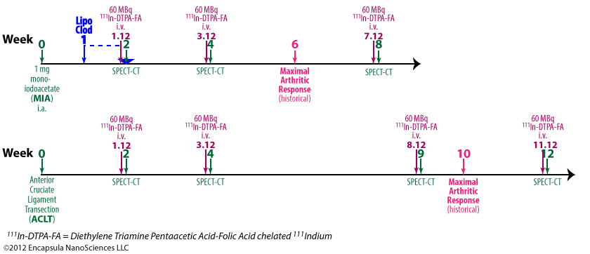
Experimental timeline

  1. Liposome prep method cited—van Rooijen N, Sanders A. Liposome mediated depletion of macrophages: mechanism of action, preparation of liposomes and applications. Journal of Immunological Methods. 1994 Sep 14;174(1–2):83–93.
  2. Rats were given intra-articular injections of clodronate liposomes in one knee while PBS was injected into the contralateral knee 7 d post-induction of arthritis in both knees.
  3. Osteoarthritis was induced by 1 mg mono-iodoacetate in 50 µl saline (MIA model).
  4. Clodronate liposome-mediated depletion was not assessed in the ACLT model.
  5. 20 h prior to SPECT-CT scans, rats were injected intravenously with 60 MBq 111In-DTPA-FA which the authors claim target activated macrophages via the folate receptor.

Results

  1. The authors report that untreated rats experience a 47% higher uptake of radiotracer in arthritic vs. non-arthritic knees.
  2. The clodronate liposome-treated knees showed 67% less radiotracer than the contralateral control arthritic knees 1 week post-injection of clodronate liposomes.
  3. Since it has been shown in mice that it takes 6-8 weeks for macrophages to fully repopulate the entire synovial lining, it would have been interesting to see subsequent (4, 6, 8 week) scans on these animals.
  4. No other experiments related to clodronate liposomes reported in this paper.


[divider_top]

2005-lawlor-3749

Lawlor KE, Wong PKK, Campbell IK, van Rooijen N, Wicks IP. Acute CD4+ T lymphocyte-dependent interleukin-1-driven arthritis selectively requires interleukin-2 and interleukin-4, joint macrophages, granulocyte-macrophage colony-stimulating factor, interleukin-6, and leukemia inhibitory factor. Arthritis & Rheumatism. 2005 Dec;52(12):3749–54.

Abstract

Objective. To further investigate the effects of interleukin-1 (IL-1) in immune-mediated joint inflammation, we examined the role of IL-2, Th1 interferon-γ (IFNγ), and Th2 (IL-4) cytokines, joint macrophages, and macrophage-derived cytokines (IL-12 p40, IL-6, leukemia inhibitory factor [LIF], oncostatin M [OSM], and granulocyte–macrophage colony-stimulating factor [GM-CSF]) in a CD4 T lymphocyte–dependent model of acute arthritis.

Methods. Methylated bovine serum albumin (mBSA)/IL-1–induced arthritis was elicited in wild-type, gene-knockout, and monoclonal antibody–treated mice. Synovial lining macrophages were selectively depleted by intraarticular injection of clodronate liposomes prior to disease induction. The severity of arthritis was assessed histologically.

Results. Mice deficient in IL-2 were almost completely protected from arthritis, and neutralization of IL-4 reduced the severity of disease. In contrast, arthritis severity and resolution appeared to be independent of IFN. Synovial lining macrophage depletion markedly reduced arthritis severity. IL-6 or LIF deficiency was only modestly protective, although as previously reported, GM-CSF deficiency conferred profound disease resistance. IL-12 p40–deficient mice (which lack IL-12 and IL-23) and OSM receptor–deficient mice were susceptible to mBSA/IL-1–induced arthritis.

Conclusion. Acute mBSA/IL-1–induced arthritis is dependent on IL-2 and IL-4, but not IFNγ. In vivo, the Th1/Th2 paradigm may be distorted by the presence of macrophage-derived cytokines such as IL-1. Synovial lining macrophages are essential in mBSA/IL-1–induced arthritis. However, the requirement for macrophage-derived cytokines is selective; that is, IL-6, LIF, and especially GM-CSF are necessary, but IL-12, IL-23, and OSM are dispensable. IL-1 may therefore influence both adaptive and innate immune mechanisms in acute inflammatory arthritis.

[custom_table]
Clodronate Concentration Total Lipid Concentration Lipid Composition Lipid Mole % Liposome Type Control Liposomes Control Free Clodronate
5 mg/ml 23.5 mg/ml EPC/chol 84/16 MLV none ND
[/custom_table]  [custom_table]
Animal Description Clodronate Dose Dosing Method/Site Target Phagocytes Systemic Dosing? Systemic Results
C57BL/6, DBA/1, male, 8-12 w 42 µg/10 µl intra-articular/knee synovial no N/A
[/custom_table]

Notes

  1. Liposome prep method cited—
    van Lent PLEM, Holthuysen AEM, van den Bersselaar LAM, van Rooijen N, Joosten LAB, van de Loo FAJ, et al. Phagocytic lining cells determine local expression of inflammation in type II collagen–induced arthritis. Arthritis & Rheumatism. 1996 Sep;39(9):1545–55.
  2. Experimental timeline

    Mice were given intra-articular injections of clodronate liposomes in one knee while PBS was injected into the contralateral knee 7 d and 4 d prior to induction of arthritis.

  3. 7 d post-first liposome treatment, at the time of maximal macrophage depletion, 2 mg in 10 µl methylated BSA was dosed intra-articularly followed by s.c. injection of 0.25 µg human IL-1β in 20 µl into the hindpaw of the same limb.
  4. The human IL-1β was dosed on days 8 and 9 as well.
  5. Mice were sacrificed 7d post-induction of arthritis and assessed histologically for disease severity.

Results

  1. The authors report a 75% reduction in histological score for disease severity in clodronate-liposome depleted animals compared to controls.
  2. No other experiments related to clodronate liposomes reported in this paper.


[divider_top]

1999-blom-1046

Blom AB, van Lent PLE, Holthuysen AE, van den Berg WB. Immune complexes, but not streptococcal cell walls or zymosan, cause chronic arthritis in mouse strains susceptible for collagen type II auto-immune arthritis. Cytokine. 1999 Dec;11(12):1046–56.

Abstract

In this study we investigated mechanisms involved in the chronic character of experimental collagen type II induced arthritis (CIA). We compared the knee joints of mouse strains which are prone to develop this autoimmune disease (DBA/1, B10RIII) with other nonsusceptible mouse strains (C57Bl/6, BALB/c) in their reaction to different stimuli: immune complexes (IC), zymosan and streptococcal cell walls (SCW). Inflammation was evaluated by 99m Tc uptake measurements and in haematoxylin- and eosin-stained knee-joint sections. Passively induced immune complex mediated arthritis (ICA) in knee joints of C57Bl/6 and BALB/c mice, showed moderate cell influx at day 3, whereas at day 7 only minor amounts of inflammatory cells were observed. In contrast, in arthritic DBA/1 and, to a lesser extent, in B10.RIII joints, a tremendous cell influx was observed at day 3 and even at day 14 there was still significant synovitis. In contrast, if arthritis was elicited by intra-articular injection of zymosan or SCW in C57Bl/6 and DBA/1, the course of inflammation was similar in both strains and no chronic inflammation developed. In line with severe arthritis, chemotactic factor production was dramatically enhanced in ICA in DBA/1 mice, and a prolonged production of IL-1 was evident. When IL-1 was neutralized before or during the ICA using specific anti-IL-1, antibodies, inflammation could be blocked completely. Single or multiple injection of IL-1 in the knee joint of C57Bl/6 or DBA/1 showed comparable inflammation, indicating that the chemotactic response per se is comparable in both strains. No prolonged production of IL-1 was found during zymosan or SCW arthritis. Selective removal of macrophages from the synovial intima prior to ICA induction (using clodronate-containing liposomes) prevented the onset of inflammation in C57Bl/6 and DBA/1 mice. It can be concluded that immune complexes, but not zymosan or SCW, cause a more severe and chronic arthritis in mouse strains which are susceptible for collagen type II autoimmune arthritis. This is due to higher and prolonged expression of IL-1 and chemotactic factors, caused by stimulation with immune complexes. The interaction of IC with lining macrophages probably plays a dominant role in development of chronicity.

[custom_table]
Clodronate Concentration Total Lipid Concentration Lipid Composition Lipid Mole % Liposome Type Control Liposomes Control Free Clodronate
12.5 mg/ml 23.1 mg/ml EPC/chol 84/16 MLV PBS ND
[/custom_table] [custom_table]
Animal Description Clodronate Dose Dosing Method/Site Target Phagocytes Systemic Dosing? Systemic Results
C57BL/6, DBA/1, male, 8-12 w 75 µg/6 µl intra-articular/knee synovial no N/A
[/custom_table]

Notes

  1. Liposome prep method cited— van Lent PL, van den Hoek AE, van den Bersselaar LA, Spanjaards MF, van Rooijen N, Dijkstra CD, et al. In vivo role of phagocytic synovial lining cells in onset of experimental arthritis. Am J Pathol. 1993 Oct;143(4):1226–37.
  2. Experimental timeline

    No explanation as to why clodronate is 2.5X more concentrated than reported in other papers. No mention of change in prep method.

  3. Mice were given intra-articular injections of clodronate liposomes in one knee while either PBS or PBS liposomes were injected into the contralateral knee.
  4. 7 d post-treatment, at the time of maximal macrophage depletion, poly-lysine-lysozyme conjugate was dosed intra-articularly 16 h post-intra-articular injection of rabbit anti-lysosome polyclonal Ab to induce immune-complex-mediated arthritis (ICA).
  5. 2 d post-induction of arthritis, mice were sacrificed for histiology or injected with 99m Tc followed by γ-counting of the knee 30 minutes later.

Results

  1. The authors reported a 57% reduction in joint swelling as measured by 99m Tc uptake in synovial macrophage-depleted C57BL/6 mice and 58% reduction in DBA/1 mice.
  2. No other experiments related to clodronate liposomes were reported in this paper.


[divider_top]

1993-vanlent-1226

van Lent PL, van den Hoek AE, van den Bersselaar LA, Spanjaards MF, van Rooijen N, Dijkstra CD, van de Putte LB, van den Ber WB. In vivo role of phagocytic synovial lining cells in onset of experimental arthritis.  Am J Pathol. 1993 Oct;143(4):1226–37.

Abstract

The in vivo role of phagocytic synovial lining cells (SLC) was studied in acute experimental arthritis in the mouse. SLCs were selectively depleted by injecting liposomes encapsulating the drug dichloromethylene diphosphonate (CL 2 MDP, clodronate). Optimal depletion of phagocytic lining cells occurred 7 days after CL 2 MDP liposome injection. Eliciting an immune complex-mediated arthritis in SLC-depleted knee joints largely prevented inflammation if compared to control arthritic knee joints. Joint swelling and influx of inflammatory cells into the joint cavity was markedly diminished. Cartilage damage, in this model related to influx of inflammatory cells, was significantly decreased. Reduced influx of inflammatory cells (mainly polymorphonuclear neutrophils) was correlated to a decreased production of chemotactic factors as measured in washouts of arthritic joints in a two-compartment Transwell system. Interleukin-1-driven chemotactic factors seem to be involved. Interleukin-1 levels were significantly lowered in SLC-depleted arthritic knee joints as compared to controls. Injection of recombinant murine interleukin-1 in SLC-depleted knee joints caused less influx of inflammatory cells as compared to injection into control knee joints. A specific damage of CL 2 MDP liposome treatment to synovial blood vessels was excluded as intraarticular injection of human recombinant C5a in lining-depleted knee joints showed similar influx of inflammatory cells if compared to human recombinant C5a injection in control knee joints. This study indicates that in immune complex-mediated arthritis, phagocytic lining cells regulate the onset of the inflammatory response.

[custom_table]
Clodronate Concentration Total Lipid Concentration Lipid Composition Lipid Mole % Liposome Type Control Liposomes Control Free Clodronate
5 mg/ml 23.5 mg/ml EPC/chol 84/16 MLV PBS ND
[/custom_table] [custom_table]
Animal Description Clodronate Dose Dosing Method/Site Target Phagocytes Systemic Dosing? Systemic Results
C57BL/6, male, 8-12 w 75 µg/6 µl intra-articular/knee eosin/hematoxylin stained no N/A
[/custom_table]

Notes

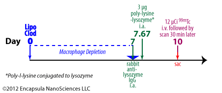
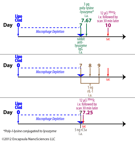
Experimental timeline for van Lent, et. al. [1993]. Liposomal clodronate or controls were injected intra-articularly on day 0 followed by induction of immune complex-mediated arithritis by various methods on day 7.

  1. Liposome prep method cited—
    van Rooijen N. The liposome-mediated macrophage “suicide” technique. J. Immunol. Methods. 1989 Nov 13;124(1):1–6.
  2. No explanation as to why clodronate is 2.5X more concentrated than reported in other papers. No mention of change in prep method.
  3. Mice were given intra-articular injections of clodronate liposomes in one knee while either PBS or PBS liposomes were injected into the contralateral knee.
  4. 7 d post-treatment, at the time of maximal macrophage depletion, poly-lysine-lysozyme conjugate was dosed intra-articularly 16 h post-intra-articular injection of rabbit anti-lysosome polyclonal Ab to induce immune-complex-mediated arthritis (ICA).
  5. 2 d post-induction of arthritis, mice were sacrificed  for histiology or injected with 99m Tc followed by γ-counting of the knee 30 minutes later.
  6. In another experiment, 1 ng rIL-1 was dosed intra-articularly for 3 days after liposome treatments. Animals were sacrificed and tissue examined for PMN infiltration.
  7. In a 3rd experiment, 5 µg human rC5a was injected into the knee joints and knees evaluated for uptake of 99mTc; histilogical analysis also performed.

Results

  1. The authors report a 67% reduction in joint swelling as measured by 99mTc uptake in synovial macrophage-depleted animals.
  2. Proteoglycan synthesis and degradation, a measure of cartilage deteriorization, was reduced by about 50% when synovial macrophages were depleted prior to arthritis induction.
  3. Cytokine washouts of arthritic joints from animals depleted of synovial macrophages was only about 50% as effective as control animal washouts in eliciting PMN migration in transwell assays.
  4. At 6 h post-induction of arthritis, the level of IL-1 measured in the synovial fluid of macrophage depleted animals was about half the level of IL-1 detected in control animal synovial fluid.
  5. Significantly fewer PMN migrated into the synovia of macrophage depleted mice post-treatment with intra-articular IL-1 compared to control animals (no cell counts reported).
  6. To ensure that clodronate liposome treatment did not adversely effect the capillaries of the synovial tissue such that inflammatory mediator effects were inhibited, human rC5a, which does not require cytokine mediators in soliciting an inflammatory response, was shown to elicit an inflammatory response irrespective of clodronate liposome treatment.
  7. 125I-labeled poly-lysine-lysozyme clearance was monitored post-intra-articular injection to confirm that the antigen complex did not leave the joint more quickly in clodronate liposome-treated animals; it did not. The level of antigen complex has been shown to correlate to arthritis severity.
  8. The authors point out that IL-1, itself, is not chemotactic, thus the mechanism of PMN infiltration is due to an indirect effect of IL-1 on inflammatory cells in initiating the release of chemotactic factors.


[divider_top]

1998-vanlent-408

van Lent PLEM, Holthuysen AEM, van Rooijen N, van de Putte LBA, van den Berg WB.  Local removal of phagocytic synovial lining cells by clodronate-liposomes decreases cartilage destruction during collagen type II arthritis. Ann Rheum Dis. 1998 Jul 1;57(7):408–13.

Abstract

OBJECTIVE: To investigate whether local removal of phagocytic synovial lining cells (SLCs) from the knee joint before onset of collagen type II arthritis has an effect on development of cartilage destruction.

METHODS: Phagocytic SLCs were selectively depleted by a single injection of clodronate laden liposomes in the knee joint seven days before induction of collagen type II arthritis (CIA). Clodronate laden liposomes were given in one knee joint either alone or in combination with a short-term oral treatment of dexamethasone. Cartilage damage including proteoglycan depletion and chondrocyte death was measured in total knee joints sections stained with safranin-o or haematoxylin.

RESULTS: Local removal of phagocytic SLCs, seven days before arthritis onset, prevented cell influx for the larger part. Chondrocyte death was significantly decreased in the SLC depleted arthritic joint both at an early (6 days) and late (12 days) time point after CIA induction. However, depletion of proteoglycans from femoral and patellar cartilage layers was not prevented. If the mild acute inflammation caused by a single clodronate laden liposome injection in the left knee joint, was blocked by a short-term (on consecutive days 9, 8, 7, 6, 5 before CIA onset) oral treatment with dexamethasone, cell influx, but also proteoglycan depletion was almost completely blocked. In the contralateral control right knee joint prominent cell influx and severe cartilage damage was observed, indicating that there was no effect of dexamethasone anymore at the onset of CIA.

CONCLUSIONS: This study shows that removal of phagocytic lining cells before CIA induction, particularly in the presence of a short-term treatment with dexamethasone, decreases cartilage destruction.

[custom_table]
Clodronate Concentration Total Lipid Concentration Lipid Composition Lipid Mole % Liposome Type Control Liposomes Control Free Clodronate
5 mg/ml 23.5 mg/ml EPC/chol 84/16 MLV PBS ND
[/custom_table]  [custom_table]
Animal Description Clodronate Dose Dosing Method/Site Target Phagocytes Systemic Dosing? Systemic Results
DBA/1 lac J, male, 10-12 w 30 µg/6 µl intra-articular/knee eosin/hematoxylin stained no N/A
[/custom_table]

Notes

    1. Liposome prep method cited—
      van Rooijen N, Sanders A. Liposome mediated depletion of macrophages: mechanism of action, preparation of liposomes and applications. Journal of Immunological Methods. 1994 Sep 14;174(1–2):83–93.
    2. Mice were given intra-articular injections of clodronate liposomes in one knee while PBS or PBS liposomes were injected into the contralateral knee.

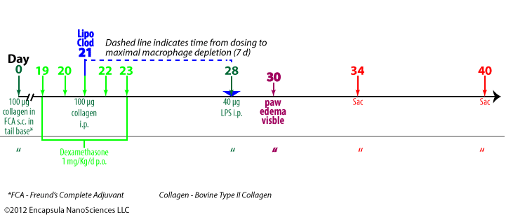
Experimental treatment timeline from van Lent, et. al., 1996. Arthritis onset is set at day 28 when the LPS is dosed. Macroscopic paw edema is visible from day 30.

  1. Mice were sacrficed at 6 d or 12 d post-onset of arthritis and joints stanined with eosin/hematoxylin (cells) or safrinin-o (proteoglycan).

Results

  1. Cartilage damage measured by proteoglycan (PG) depletion was similar in all animals despite effective synovial lining cell (SLC) depletion.
  2. Chrondrocytes are constituent cells of the cartilage that synthesize and degrade cartilage components.
  3. At 6 d post-onset, chondrocyte death in the femur was reduced by 25% in the femur and 90% in the patella and at 12 d, chondrocyte death was reduced by ~75% in the femur and about 50% in the patella.
  4. When dexamethasone treatment is combined with clodronate liposome treatment, PG depletion is also inhibited by 75-80%.


[divider_top]

1996-vanlent-1545

van Lent PLEM, Holthuysen AEM, van den Bersselaar LAM, van Rooijen N, Joosten LAB, van de Loo FAJ, van de Putte LBA, van den Berg WB.  Phagocytic lining cells determine local expression of inflammation in type II collagen–induced arthritis.Arthritis & Rheumatism. 1996 Sep;39(9):1545–55.

Abstract

Objective . To investigate the in vivo role of phagocytic synovial lining cells in the local expression of inflammation in type II collagen–induced arthritis (CIA) in DBA/1J mice.

Methods . On various days before arthritis induction (day 7, 5, or 2), phagocytic lining cells were selectively depleted from the synovial layer by injecting multilamellar liposomes containing clodronate (dichloromethylene diphosphonate) directly into the knee joints. As controls, either PBS or PBS-laden liposomes were injected. CIA was induced by immunizing mice with heterologous bovine type II collagen in Freund’s complete adjuvant. Arthritis onset was synchronized by a single intraperitoneal injection of lipopolysaccharide; arthritis was evaluated in hematoxylin and eosin–stained knee joint sections. Chemotactic activity in synovial washout samples was detected in a Transwell chemotactic assay. Interleukin-1 (IL-1) and tumor necrosis factor α (TNFα) protein levels were measured in NOB-1 and L929 bioassays, respectively. IL-1 messenger RNA (mRNA) in synovial specimens was measured by reverse transcriptase–polymerase chain reaction. IL-1 was also detected immunohistologically in knee joint sections.

Results . In clodronate-laden liposome–treated, lining-depleted knee joints, there was significantly decreased inflammation compared with controls. Cell influx into the synovium was markedly decreased. Expression of IL-1 mRNA in the synovium was significantly reduced. IL-1 was detected only in some cells in the deeper synovial layer, in contrast to controls, in which large numbers of cells in the deeper synovial layer were stained. In synovial washouts from lining-depleted knee joints, biologically active IL-1 levels were reduced by 40% at 6 hours after arthritis induction. Most strikingly, chemotactic activity was highly decreased in these synovial washout samples. When IL-1 or TNFα was injected into the knee joints of immunized mice in which arthritis was not yet expressed, arthritis was not induced in the lining-depleted joints, whereas marked cell influx was found in control joints.

Conclusion . Our data indicate that phagocytic lining cells play a crucial role in the local expression of inflammation in systemically induced CIA. Phagocytic lining cells probably form an important source of chemotactic factors which are set free upon activation by IL-1 or TNFα.

[custom_table]
Clodronate Concentration Total Lipid Concentration Lipid Composition Lipid Mole % Liposome Type Control Liposomes Control Free Clodronate
5 mg/ml 23.5 mg/ml EPC/chol 84/16 MLV PBS ND
[/custom_table] [custom_table]
Animal Description Clodronate Dose Dosing Method/Site Target Phagocytes Systemic Dosing? Systemic Results
DBA/1 lac J, male, 10-12 w 30 µg/6 µl intra-articular/knee eosin/hematoxylin stained no N/A
[/custom_table]

Notes

  1. Liposome prep method cited— van Rooijen N, Sanders A. Liposome mediated depletion of macrophages: mechanism of action, preparation of liposomes and applications. Journal of Immunological Methods. 1994 Sep 14;174(1–2):83–93.
  2. Mice were given intra-articular injections of clodronate liposomes in one knee while either PBS or PBS liposomes were injected into the contralateral knee.

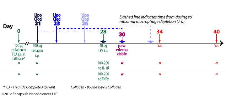
    Experimental treatment protocol described in van Lent, et. al. , 1996. Macroscopic paw edema was visible by 2 days post-onset of arthritis (day 28). In separate experiments either IL-1β or TNFα instead of LPS was used to initiate arthritis on day 28.

  3. Hind paws became visibly swollen 2 days post-onset of arthritis at day 28.
  4. Mice were sacrificed either 6 days or 12 days post-onset of arthritis at day 28.

Results

  1. The total number of cells found in synovial fluid (exudate) did not decrease significantly from control values in arthritic animals that were treated with intra-articular liposomal clodronate either 7, 5 or 2 d prior to the onset of arthritis. The animals were sacrificed 6 d post-onset and the cell count (eosin/hematoxylin stained) was determined microscopically.
  2. The total number of cells found in synovial layer (infiltrate) in liposomal-clodronate-treated animals was only about 20-30% of the total cells found in control (PBS or PBS liposomes) animals.
  3. The synovial layer of liposomal clodronate treated animals showed about 75% reduction in both neutrophils and macrophages, although the ratio of macrophages to neutrophils did not change.
  4. The effect was observed only in the treated knee and not the contralateral knee of an animal and no differences were seen between animals that were treated at differing times before onset.
  5. Clodronate liposome treatment had no effect on hindpaw swelling of the treated limb.
  6. Similar responses were seen in animals that were sacrificed 12 days post-onset of arthritis.
  7. Synovial washouts of treated animals (7 d prior to onset) either 6 h or 48 h post-onset of arthritis showed a 70% (6 h) and 95% (48 h) reduction in their ability to stimulate migration of PMN.
  8. At 6 h post-onset, IL-1 β mRNA levels were reduced 4X in animals treated 7 d post-onset with clodronate liposomes compared to controls.
  9. At 6 h post-onset, IL-1 β protein level was reduced by about 40% in liposome treated animals compared to controls. At 2 d post-onset, IL-1 β was minimal in all groups.
  10. Clodronate liposome treatment completely abolished cell-influx (counted 2 d post-onset) into joints treated with either 100 or 200 ng IL-1 β or 100 or 200 ng TNF α injected on day 28 (instead of LPS).
  11. The authors suggested that they thought that clodronate-liposome mediated depletion of macrophages may enhance the arthritic response due to cytokine release as a result of macrophage death. Since this was not the case, they speculate that either cytokine release is minimal during depletion or the cytokines may have been contained in apoptotic bodies.
  12. The authors conclude that the cells of the synovial lining are the key sources of cytokine-induced influx of inflammatory cells post-onset of collagen-induced arthritis.


[divider_top]

2000-vanderhoek-1059

van der Hoek KH, Maddocks S, Woodhouse CM, van Rooijen N, Robertson SA, Norman RJ.
Intrabursal Injection of Clodronate Liposomes Causes Macrophage Depletion and Inhibits Ovulation in the Mouse Ovary.
Biology of Reproduction. 2000 Apr 1;62(4):1059 –1066.
[toggle_content title=”Abstract”]To investigate the role of the ovarian macrophage population in ovulation, we examined the effect of depleting this population using liposome-encapsulated clodronate. Clodronate liposomes, saline liposomes, or saline alone was injected under the ovarian bursa in gonadotropin-primed adult mice, either 84 h (Day −3) or 36 h (Day −1) before ovulation. Ovulation rates were determined by counting the number of oocytes released. The numbers of graafian follicles and corpora lutea were also counted immediately before and after ovulation. Macrophage distribution within the theca and stroma of preovulatory ovaries was examined by immunohistochemistry with specific monoclonal antibodies to the macrophage antigens macrosialin, major histocompatability complex class II (Ia), and F4/80. Injection of clodronate liposomes on Day −1 did not affect ovulation rates, whereas administration on Day −3 caused a significant reduction in ovulation rate (mean oocytes ovulated = 5.25 ± 0.6 from clodronate liposome-treated ovaries and 9.13 ± 0.9 from saline-treated ovaries, respectively, P < 0.05). The numbers of macrosialin-positive macrophages present in the theca at ovulation were reduced by treatment with clodronate liposomes on Day −1, and treatment on Day −3 reduced the numbers of Ia-positive and macrosialin-positive macrophages present in the theca. When the subsequent ovarian cycles were examined by vaginal smearing, the metestrous-2/diestrous stage was found to be extended in clodronate liposome-treated animals (7.5 ± 1.3 days vs. 3.4 ± 0.4 days for saline liposome-treated animals, P < 0.05). These results suggest that thecal macrophages may be involved in the regulation of follicular growth and rupture, as well as being important for the normal progression of the estrous cycle.[/toggle_content] [toggle_content title=”Clodronate Liposome Parameters”] [custom_table]
Clodronate Concentration Total Lipid Concentration Lipid Composition Lipid Mole % Liposome Type Control Liposomes Control Free Clodronate
5 mg/ml 23.5 mg/ml EPC/Chol 84/16 MLV Saline ND
[/custom_table] [/toggle_content] [toggle_content title=”Animals and Dosing”] [custom_table]
Animal Description Clodronate Dose Dosing Method/Site Target Phagocytes Systemic Dosing? Systemic Results
C57BL/61 mice, female, 8-11 w 10 µl intra-bursal/ovary F4/80+FA/11+MHC II (Ia)+ macrophages no N/A

1Paper states that C57B16 mice were used; we assume that this is an error.

[/custom_table] [/toggle_content] [toggle_content title=”Notes”]
  1. Liposome prep method cited—
    van Rooijen N, Sanders A. Liposome mediated depletion of macrophages: mechanism of action, preparation of liposomes and applications. Journal of Immunological Methods. 1994 Sep 14;174(1–2):83–93.
[/toggle_content] [toggle_content title=”Results”]
  1. Intrabursally-injected fluorescent liposomes were confirmed to enter the theca and stroma of the ovary, but not the interior sections of the ovary. Unfortunately, the cellular localization of these liposomes was not determined, however the fluorescence signal decrease to baseline by 6 h. Therefore, we assume that the liposomes were phagocytosed and metabolized, but the cellular distribution of the liposomes would verify that all macrophages were accessible by liposomes.
  2. Macrophage depletion was only about 30% at best and varied dramatically depending on macrophage phenotype and tissue location. If a cellular distribution of fluorescent liposomes had been determined, the reason behind the limited depletion may have been clear.
  3. Clodronate liposomes, but not control liposomes, reduced the ovulation rate by 50-60% when the clodronate liposomes were dosed 3 days, but not 1 day, prior to induction of ovulation.
  4. The authors cite the possibility that rapid repopulation of ovarian macrophages may be a reason for incomplete depletion of the population. If this were true, additional systemic dosing should deplete circulating monocytes from which the ovarian macrophages mature, thus limiting the influx of newly recruited monocytes/macrophages.
[/toggle_content]
[divider_top]

1994-vanlent-221

van Lent PLEM, van den Bersselaar LAM, Holthuyzen AEM, van Rooijen N, van de Putte LBA, van den Berg WB.
Phagocytic synovial lining cells in experimentally induced chronic arthritis: down-regulation of synovitis by CL2MDP-liposomes.
Rheumatology International. 1994;13(6):221–8.
[toggle_content title=”Abstract”]Chronic inflammation of the joint is characterized by the long-term presence of macrophage-like cells in the multilayered synovium. We examined whether synovial phagocytic cells which have settled in the inflamed lining layer play a role in perpetuating synovitis by selectively eliminating them from chronically arthritic murine knee joints. For this purpose we used liposomes encapsulating the drug dichloromethylene diphosphonate (CL2MDP, Clodronate). Injection of CL2MDP-liposomes into acutely inflamed knee joints (6h, 1 and 3 days) had no significant effect on late chronic synovitis (14 and 21 days after arthritis induction) as observed in haematoxylin and eosin-stained total knee joint sections. Liposomes did not reach the lining layer, as seen with fluorescent liposomes. Additional in vitro studies revealed that activated polymorphs were not affected by CL2MDP-liposomes within 16 h of incubation. Liposomes formed clusters, however, in the presence of intact polymorphs or extracts of polymorphs. In contrast, a significant down-regulation of late synovitis was observed if CL2MDP-liposomes were given during the chronic phase (day 7). Phosphate-buffered saline (PBS) alone or PBS-liposomes had no effect on synovitis. A single injection of CL2MDP-liposomes eliminated many of the phagocytic lining cells and deeper lying inflammatory cells for at least 4 weeks. Free CL2MDP had a minor but significant effect. This study indicates that phagocytic synovial lining cells play an important role in propagating chronic synovitis. To eliminate them from inflamed knee joints, CL2MDP-liposomes should be injected in the chronic and not in the early arthritic phase.[/toggle_content] [toggle_content title=”Clodronate Liposome Parameters”] [custom_table]
Clodronate Concentration Total Lipid Concentration Lipid Composition Lipid Mole % Liposome Type Control Liposomes Control Free Clodronate
12.5 mg/ml1 23.5 mg/ml EPC/Chol 84/16 MLV PBS ND

1This number is very high for the amount of lipid and the prep method cited. Typically this prep would contain around 5 mg/ml clodronate.

[/custom_table] [/toggle_content] [toggle_content title=”Animals and Dosing”] [custom_table]
Animal Description Clodronate Dose Dosing Method/Site Target Phagocytes Systemic Dosing? Systemic Results
C57BL/6 mice, male, 8-12 w, 25-30 g 75 µg/6 µl intra-articular/knee synovial no N/A
[/custom_table] [/toggle_content] [toggle_content title=”Notes”]
  1. Liposome prep method cited—
    van Rooijen N. The liposome-mediated macrophage “suicide” technique. J. Immunol. Methods. 1989 Nov 13;124(1):1–6.
  2. As stated above, this clodronate concentration in the liposomes is about 2.5 higher than that usually reported. The authors do not report measuring the clodronate and lipid concentrations in the final prep
[/toggle_content] [toggle_content title=”Results”]
  1. When when a single dose of clodronate liposomes was administered 7 d, but not 0.25, 1 or 3 d, post-induction of arthritis, synovitis was alleviated for at least 4 weeks.
  2. A single dose of free clodronate at 7 d also provided a significant reduction in synovitis, but was only about 50% as effective as liposomal clodronate.
  3. When fluorescent clodronate liposomes were injected at the earlier time points (0.25, 1, 3 d), the liposomes did not localize to the phagocytic cells of the synovial lining, while liposomes injected at 7 d post-induction of arthritis were almost exclusively found in the phagocytes.
  4. When incubated in vitro (5% FCS), clodronate liposomes and activated neutrophils aggregate; free clodronate nor PBS causes any aggregation and control liposomes cause slight aggregation under similar conditions.
    • Do the clodronate liposomes alone aggregate in the incubation media?
    • Does the incubation/aggregation result in release of clodronate from the liposomes?
    • Would a mixture of free clodronate and control liposomes result in aggregation?
    • How similar in size and morphology are the clodrosome and control liposomes?
    • Would clodronate liposomes which were pre-incubated in serum cause neutrophil aggregation?
    • Would the aggregation occur in serum-free media?
    • Would clodronate liposomes which were pre-incubated in serum cause neutrophil aggregation in serum-free media?

    Liposomes are known to aggregate in serum containing media and some believe that the mechanism of enhanced circulation times of small liposomes containing PEG lipids is related to the fact that the pegylated liposomes do not aggregate in serum. As to the authors’ speculation that this aggregation in the presence of  neutrophils prevents liposomes from interacting with the phagocytes in the acute phase of arthritis, this question could be easily answered.

    • Since this aggregation phenomenon must be a saturable process, multiple injections of clodronate liposomes timed closely together will saturate the binding sites on the neutrophils and/or temporarily deplete serum components which contribute to the aggregation. Once saturation is complete, clodronate liposomes will be available to the phagocytes.
    • The difference in behaviors between the clodronate and control liposomes is less clear, however we know that it is not uncommon for a drug-laden liposome to have a different size distribution and morphology when compared to “empty” liposomes of the same lipid composition. This factor may be at work here.
  5. Neutrophil lysates cause aggregation and destruction of liposomes; not surprising since phospholipases are among the most predominant destructive enzymes in granules.
  6. Under the same incubation conditions, activated neutrophils die at about 4 hours independent of the additive (clodronate liposomes, free clodronate, control liposomes, PBS).
  7. Clodronate liposomes did not affect Ag removal from the synovial fluid.
[/toggle_content]
[divider_top]