Piscaer TM, Müller C, Mindt TL, Lubberts E, Verhaar JAN, Krenning EP, Shibli R, de Jong M, Weinans H. Imaging of activated macrophages in experimental osteoarthritis using folate-targeted animal single-photon-emission computed tomography/computed tomography. Arthritis & Rheumatism. 2011 Jul;63(7):1898–907.
Abstract
Objective. Evaluation of macrophage activation may provide essential information about aetiology and progression rate of osteoarthritis. Activated macrophages abundantly express the folate-receptor-beta (FR-β), which can be targeted using radioactive labelled folic acid. The purpose of this study was to investigate if macrophage activation can be monitored in small animal OA models using a folate radiotracer and to test if macrophage activation differs in different models of OA and subsequent different OA progression.
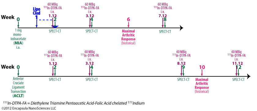
Methods. Two rat models of OA were used: the monoiodoacetate (MIA) model, which is a fast progressing biochemical induced model and the anterior cruciate ligament transaction (ACLT) model that induces OA at a slower pace. Images were obtained using high resolution small animal SPECT/CT. Specificity of the technique was tested by eradicating macrophages using clodronate laden liposomes and blockade of the FR-β by cold folic acid.
Results. The MIA model had a high initial activation with a peak after two weeks which disappeared after eight weeks. The ACLT model showed less activation but was still active 12 weeks after induction. The technique allowed monitoring of the disease process over time, in which late stage disease showed less macrophage activation than early onset stages especially in the fast progressing MIA model for OA.
Conclusion. Macrophage activation in experimental OA could clearly be demonstrated and monitored by the folate radiotracer. The high resolution, high sensitivity and high specificity of the used technique allowed clear localisation of macrophage activity in a disease model, which is not known for abundant macrophage involvement.
[custom_table]| Clodronate Concentration | Total Lipid Concentration | Lipid Composition | Lipid Mole % | Liposome Type | Control Liposomes | Control Free Clodronate |
|---|---|---|---|---|---|---|
| 5 mg/ml | 23.5 mg/ml | EPC/chol | 84/16 | MLV | none | ND |
| Animal Description | Clodronate Dose | Dosing Method/Site | Target Phagocytes | Systemic Dosing? | Systemic Results |
|---|---|---|---|---|---|
| Wistar rats, male, 14 w | 50 µl | intra-articular/knee | synovial MΦ | no | N/A |
Notes
- Liposome prep method cited—van Rooijen N, Sanders A. Liposome mediated depletion of macrophages: mechanism of action, preparation of liposomes and applications. Journal of Immunological Methods. 1994 Sep 14;174(1–2):83–93.
- Rats were given intra-articular injections of clodronate liposomes in one knee while PBS was injected into the contralateral knee 7 d post-induction of arthritis in both knees.
- Osteoarthritis was induced by 1 mg mono-iodoacetate in 50 µl saline (MIA model).
- Clodronate liposome-mediated depletion was not assessed in the ACLT model.
- 20 h prior to SPECT-CT scans, rats were injected intravenously with 60 MBq 111In-DTPA-FA which the authors claim target activated macrophages via the folate receptor.
Results
- The authors report that untreated rats experience a 47% higher uptake of radiotracer in arthritic vs. non-arthritic knees.
- The clodronate liposome-treated knees showed 67% less radiotracer than the contralateral control arthritic knees 1 week post-injection of clodronate liposomes.
- Since it has been shown in mice that it takes 6-8 weeks for macrophages to fully repopulate the entire synovial lining, it would have been interesting to see subsequent (4, 6, 8 week) scans on these animals.
- No other experiments related to clodronate liposomes reported in this paper.
